


NEWS
Health updates and
clinic news
HEPATIC HEMANGIOMA: A COMMON BENIGN LIVER TUMOR
News
Oct 12, 2026
Overview
A hepatic hemangioma (also known as a liver hemangioma) is the most common type of benign liver tumor. It is a non-cancerous growth formed by an abnormal cluster of blood vessels. In the vast majority of cases, these tumors are harmless, do not cause symptoms, and do not require active treatment.
Statistics show that hepatic hemangiomas affect approximately 0.4% to 7.3% of the population. They are more prevalent in women, particularly between the ages of 30 and 50. Most cases are incidental findings, discovered by chance during a routine abdominal ultrasound.
Causes and Risk Factors
To date, the exact cause of hepatic hemangiomas remains unknown. However, several factors are closely linked to their development:
- Gender: They are more common in women, suggesting a role for estrogen.
- Hormonal Factors: Pregnancy or the use of estrogen-containing oral contraceptives may trigger the tumor to grow faster.
- Congenital Origin: Some cases are believed to be present from birth as a vascular malformation.
- Pre-existing Liver Conditions: Often detected in patients with chronic liver disease, though they are not the direct cause.
Clinical Symptoms
The majority of patients have no symptoms. About 70–80% of hemangiomas are found incidentally during ultrasound or CT scans.
When a tumor exceeds 5 cm (known as a giant hepatic hemangioma), symptoms may include:
- Pain or discomfort in the Right Upper Quadrant (RUQ).
- A sensation of fullness, bloating, or indigestion.
- Rare cases: Nausea, loss of appetite, or unexplained weight loss.
Rare but Serious Complications:
- Spontaneous Rupture: Occurs in less than 1% of cases, causing internal bleeding that requires emergency medical attention.
- Compression: Extremely large masses may press against bile ducts or the portal vein.
Diagnosis
- The most common first-line screening method.
- Typical findings show a well-defined, hyperechoic (bright) mass.
2. Contrast-Enhanced CT Scan
- Essential for ruling out malignancy.
- Classic Sign: The tumor shows strong enhancement in the arterial phase, followed by a slow wash-out in later phases.
3. Magnetic Resonance Imaging (MRI)
- Provides higher accuracy than CT, especially for atypical or very small nodules.
- It is often the preferred choice when other imaging results are inconclusive.
- Liver function tests (AST, ALT) and bilirubin levels are usually normal.
- Tumor markers (AFP, CEA, CA 19-9) do not increase in patients with a simple hemangioma.
Note: Diagnosis is based on imaging findings. A liver biopsy is strictly avoided due to the high risk of hemorrhage (severe bleeding) from the vascular mass.
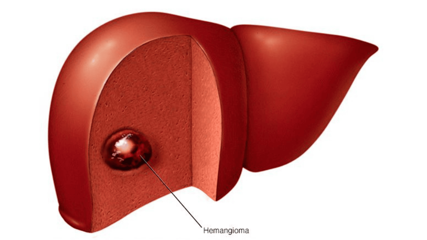
Diagnostic Criteria for Hepatic Hemangioma
A liver mass is diagnosed as a hepatic hemangioma when it meets the following criteria:
- Size: Variable, typically less than 5 cm.
- Ultrasound: A well-defined, homogeneous hyperechoic mass.
- Contrast-enhanced CT/MRI: Peripheral enhancement in the arterial phase, followed by gradual centripetal filling in delayed phases.
- No malignancy: Absence of vascular invasion or metastasis.
- Blood tests: All liver functions and tumor markers are within normal limits.
Treatment and Management
1. Asymptomatic Cases
- Monitoring: No active treatment is required.
- Follow-up: Periodic ultrasounds every 6–12 months for the first 1–2 years to ensure the tumor is stable.
2. Symptomatic or Giant Hemangiomas
Treatment is only considered if:
- The tumor is > 5 cm and causes persistent pain or discomfort.
- The tumor shows rapid growth.
- Complications such as rupture or organ compression occur.
Intervention Options:
- Surgical Resection: The primary choice for giant hemangiomas with severe symptoms.
- Transarterial Embolization (TAE): Blocking the blood supply to shrink the tumor.
- Radiofrequency Ablation (RFA): A minimally invasive option used in specific cases.
3. No Role for Medication
- There is no medication proven to shrink a liver hemangioma.
- Patients should avoid unverified herbal remedies that may cause liver toxicity.

Prevention and Lifestyle
- Adhere to Follow-ups: Stay on track with the imaging schedule recommended by your specialist.
- Hormone Consultation: Women planning a pregnancy or using hormonal therapy should discuss monitoring with their doctor.
- Liver Health: Limit alcohol intake and maintain a healthy weight to protect overall liver function.
Prognosis of Hepatic Hemangioma
- Benign Nature: Hepatic hemangiomas are non-cancerous; in most cases, they do not impact life expectancy or overall quality of life.
- Low Risk: The probability of developing complications is extremely low.
- Positive Outlook: The prognosis is excellent, requiring only periodic monitoring and routine follow-ups
FAQ
1. Is a liver hemangioma a form of cancer?
No. It is a benign growth and does not transform into cancer.
2. Does it always require surgery?
No. Most patients live their entire lives without needing surgery. It is only reserved for cases with severe symptoms or complications.
3. Will it disappear on its own?
Generally, no. They tend to remain stable in size for many years.
4. Can pregnancy make the tumor grow?
Yes. Due to the surge in estrogen, the tumor may grow faster during pregnancy. Close monitoring is advised.
5. Do I need to follow a special diet?
No specific dietary restrictions are required. A balanced, healthy diet is always recommended for general liver health.
To schedule a consultation and monitoring for Hepatic Hemangioma at DYM Medical Center Vietnam, please contact:
-
DYM Medical Center Saigon: Room B101-B103, Basement 1, mPlaza Saigon Building, No. 39 Le Duan, Sai Gon Ward, Ho Chi Minh City
-
DYM Medical Center Phu My Hung: Room 3A01, 3A Floor, The Grace Building, 71 Hoang Van Thai Street, Tan My Ward, Ho Chi Minh City
-
DYM Medical Center Hanoi: B1 Basement, Epic Tower, Alley 19 Duy Tan, Cau Giay Ward, Ha Noi
-
Hotline: 1900 29 29 37
Register now to receive the latest offers
Book an appointmentMedical specialties
- Corporate health check-up
- Dental Checkup Package
- Pediatric Examination Package
- Work Permit Examination Package - Work Permit Supplement
- Pre-wedding health check-up
- Obstetrics and Gynecology Package
- Cancer Screening Package
- Health check-up for individuals
- Japan pre-entry Tuberculosis Screening(Jpets)
- Food Handler package
- Medical examination for labor export to Japan
- Medical examination for specific skills to work in Japan (Tokutei)








Contact us
Reach out to DYM for quick support through
our official channels.